Topic 1 - Cell Biology
1.1 Introduction To Cells
UNDERSTANDINGS, APPLICATIONS AND SKILLS:
1.1.U1 According to the cell theory, living organisms are composed of cells.
1.1.U2 Organisms consisting of only one cell carry out all functions of life in that cell. [Students are expected to be able to name and briefly explain these functions of life: nutrition, metabolism, growth, response, excretion, homeostasis and reproduction.]
1.1.U3 Surface area to volume ratio is important in the limitation of cell size.
1.1.U4 Multicellular organisms have properties that emerge from the interaction of their cellular components.
1.1.U5 Specialised tissues can develop by cell differentiation in multicellular organisms.
1.1.U6 Differentiation involves the expression of some genes and not others in a cell’s genome.
1.1.U7 The capacity of stem cells to divide and differentiate along different pathways is necessary in embryonic development and also makes stem cells suitable for therapeutic uses.
1.1.A1 Questioning the cell theory using atypical examples, including striated muscle, giant algae and aseptate fungal hyphae.
1.1.A2 Investigation of functions of life in Paramecium and one named photosynthetic unicellular organism. [ Chlorella or Scenedesmus are suitable photosynthetic unicells, but Euglena should be avoided as it can feed heterotrophically.]
1.1.A3 Use of stem cells to treat Stargardt’s disease and one other named condition.
1.1.A4 Ethics of the therapeutic use of stem cells from specially created embryos, from the umbilical cord blood of a new-born baby and from an adult’s own tissues.
1.1.S1Use of a light microscope to investigate the structure of cells and tissues, with drawing of cells. Calculation of the magnification of drawings and the actual size of structures and ultrastructures shown in drawings or micrographs. (Practical 1) [ Scale bars are useful as a way of indicating actual sizes in drawings and micrographs.]
CELL THEORY
Questioning the cell theory using atypical examples:
SURFACE AREA TO VOLUME RATIO
Metabolic reactions happen in the cytoplasm of the cell and for this to continue in the cell, substances used in the reactions must be absorbed by the cell and waste products must be removed. Substances move into and out of the cell through the plasma membrane. The rate and amount of which these substances cross through the membrane depend on the surface area. Therefore, surface are to volume ratio of the cell is important. If the ration is too small (In cm (SA = 384 V = 512 SA/V ratio =0.75) is smaller then (SA = 24 V = 8 SA/V ratio = 3.0) ratio) then substances will not enter the cell as quickly as they are required and waste products will accumulate because they are produced more rapidly then they can be excreted. It is also important in relation to heat production and loss. If the ratio is too small then the cell may overheat because the metabolism produces heat faster then it is lost over the cell's surface.
MULTICELLULAR ORGANISMS AND THEIR EMERGENT PROPERTIES
CELL DIFFERENTIATION IN MULTICELLULAR ORGANISMS
GENE EXPRESSION AND CELL DIFFERENTIATION
STEM CELLS
Key properties:
APPLICATION:
Use of stem cells to treat Stargardt’s disease and one other named condition.
Therapeutic uses
APPLICATION:
Ethics of the therapeutic use of stem cells from specially created embryos, from the umbilical cord blood of a new-born baby and from an adult’s own tissues.
Ethical Considerations of Using Stem Cells
- Cells are the fundamental building blocks of ALL living organisms
- Cells are the SMALLEST organisms of life
- ALL cells arise from pre-existing cells
Questioning the cell theory using atypical examples:
- Striated muscle - The building blocks of this are not cells but muscle fibres, similar to cells. They have up to several hundred nucleuses.
- Aseptate Fungi - Consist of thread-like structures called hyphae, usually divided by septa however in aseptate fungi they don't have that.
- Giant Alga - Consists of only one, big cell.
- M - metabolism - chemical reactions inside of the cell, including cell respiration to release energy
- R - reproduction - producing offspring either sexually or asexually
- H - homeostasis - keeping conditions inside the organism within tolerable limits (temperature, water levels ....)
- G - growth - an irreversible increase in size
- R - response - the ability to react to changes in the envirnment
- E - excretion - getting rid of the waste products of metabolism
- N - nutrition - obtaining food, to provide energy and the materials needed for growth
SURFACE AREA TO VOLUME RATIO
Metabolic reactions happen in the cytoplasm of the cell and for this to continue in the cell, substances used in the reactions must be absorbed by the cell and waste products must be removed. Substances move into and out of the cell through the plasma membrane. The rate and amount of which these substances cross through the membrane depend on the surface area. Therefore, surface are to volume ratio of the cell is important. If the ration is too small (In cm (SA = 384 V = 512 SA/V ratio =0.75) is smaller then (SA = 24 V = 8 SA/V ratio = 3.0) ratio) then substances will not enter the cell as quickly as they are required and waste products will accumulate because they are produced more rapidly then they can be excreted. It is also important in relation to heat production and loss. If the ratio is too small then the cell may overheat because the metabolism produces heat faster then it is lost over the cell's surface.
MULTICELLULAR ORGANISMS AND THEIR EMERGENT PROPERTIES
- Organisms consisting of a single mass of cells, fused together, are multicellular.
- Multicellular organisms can be regarded as cooperative groups, without any cells in the group acting as a leader or supervisor.
- The characteristics of the whole organism, including the fact that it is alive, are known as emergent properties.
- Emergent properties arise from the interaction of component parts of a complex structure.
- This is summed up in the phrase: "the whole is greater then the sum of it's parts".
CELL DIFFERENTIATION IN MULTICELLULAR ORGANISMS
- In multicellular organisms different cells perform different functions, this is called 'division of labour'.
- Often a group of cells specialise in the same way to perform the same function. They are called tissue.
- The development of cells in different ways to carry out specific functions is called differentiation.
GENE EXPRESSION AND CELL DIFFERENTIATION
- All different cell types have the same set of genes.
- There are around 25,000 genes in the human genome but in most cell types less then half of the genes will be needed or used.
- A gene that is being used is being "expressed". Therefore, the gene is switched on and the information in it is being used to make a protein or other gene products.
- Cell differentiation happens because a different sequence of genes is being expressed in differential types.
- Therefore the control of gene expression is the key to development.
STEM CELLS
Key properties:
- Divide continuously, again and again.
- Not fully differentiated
- Animal life starts when the sperm fertilises the egg, which produces a zygote.
- When the zygote divides, it produces a two cell embryo.
- This two cell embryo divides and divides again and again to produce a four cell embryo, eight cell embryo, sixteen cell embryo and so on.
- At this early stage the cells are capable of dividing many times to produce large amounts of tissue.
- These cells are extremely versatile and can differentiate along many different pathways into any cell types.
- The name stem cell was given to the zygote and the cells of the early embryo, meaning all the tissues of the adult stem from them.
- Embryonic
- Cord Blood
- Adult
APPLICATION:
Use of stem cells to treat Stargardt’s disease and one other named condition.
Therapeutic uses
APPLICATION:
Ethics of the therapeutic use of stem cells from specially created embryos, from the umbilical cord blood of a new-born baby and from an adult’s own tissues.
Ethical Considerations of Using Stem Cells
|
|
|
|
1.2 Ultrastructure Of Cells
UNDERSTANDINGS, APPLICATIONS AND SKILLS:
1.2.U1Prokaryotes have a simple cell structure without compartmentalization.
1.2.U2Eukaryotes have a compartmentalized cell structure.
1.2.U3Electron microscopes have a much higher resolution than light microscopes.
1.2.A1Structure and function of organelles within exocrine gland cells of the pancreas and within palisade mesophyll cells of the leaf.
1.2.A2Prokaryotes divide by binary fission.
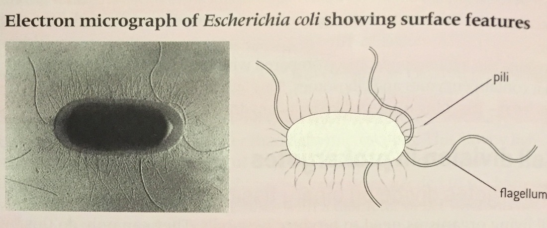
1.2.S1Drawing of the ultrastructure of prokaryotic cells based on electron micrographs.[Drawings of prokaryotic cells should show the cell wall, pili and flagella, and plasma membrane enclosing cytoplasm that contains 70S ribosomes and a nucleoid with naked DNA.]
1.2.S2Drawing of the ultrastructure of eukaryotic cells based on electron micrographs.[Drawings of eukaryotic cells should show a plasma membrane enclosing cytoplasm that contains 80S ribosomes and a nucleus, mitochondria and other membrane-bound organelles are present in the cytoplasm. Some eukaryotic cells have a cell wall.]
1.2.S3Interpretation of electron micrographs to identify organelles and deduce the function of specialized cells.
1.2.U1Prokaryotes have a simple cell structure without compartmentalization.
1.2.U2Eukaryotes have a compartmentalized cell structure.
1.2.U3Electron microscopes have a much higher resolution than light microscopes.
1.2.A1Structure and function of organelles within exocrine gland cells of the pancreas and within palisade mesophyll cells of the leaf.
1.2.A2Prokaryotes divide by binary fission.
1.2.S1Drawing of the ultrastructure of prokaryotic cells based on electron micrographs.[Drawings of prokaryotic cells should show the cell wall, pili and flagella, and plasma membrane enclosing cytoplasm that contains 70S ribosomes and a nucleoid with naked DNA.]
1.2.S2Drawing of the ultrastructure of eukaryotic cells based on electron micrographs.[Drawings of eukaryotic cells should show a plasma membrane enclosing cytoplasm that contains 80S ribosomes and a nucleus, mitochondria and other membrane-bound organelles are present in the cytoplasm. Some eukaryotic cells have a cell wall.]
1.2.S3Interpretation of electron micrographs to identify organelles and deduce the function of specialized cells.
Microscopes
Electron microscopes have a much higher resolution than light microscopes or our eyes.
Prokaryotes
Things to know:
How to draw a prokaryotic cell, Escherichia Coli, based on an electron micrograph:
Must show - Cell wall, Pili, Flagella, Ribosomes, Plasma membrane, Cytoplasm, Nucleoid region.
Electron microscopes have a much higher resolution than light microscopes or our eyes.
Prokaryotes
Things to know:
- They have a simple structure without compartments.
- They don't have a nucleus .
- The interior is entirely filled with cytoplasm. Part of which appears lighter then the rest on electron micrographs. This is because these regions contain the DNA of the cell in the form of one circular DNA molecule. This DNA is not associated with proteins, explaining the lighter appearance. This lighter area is called the nucleic, which means nucleus like, as it contains DNA but is not a true nucleus.
- They don't have cytoplasmic organelles apart from ribosomes
- Prokaryotic cells divide by binary fission and is used for asexual reproduction.
- Each of the daughter cells contains one copy of the chromosome so they are genetically identical.
- The single circular chromosome is replicated.
- The two copies of the chromosome move to opposite ends of the cell.
- Division of the cytoplasm of the cell follows.
How to draw a prokaryotic cell, Escherichia Coli, based on an electron micrograph:
Must show - Cell wall, Pili, Flagella, Ribosomes, Plasma membrane, Cytoplasm, Nucleoid region.

|

|
Eukaryotes
Things to know:
How to draw a eukaryotic cell, based on an electron micrograph:
Must show - Plasma membrane, Cytoplasm with 80S ribosomes, Nucleus, Mitochondria, and all other organelles in the cell
Things to know:
- Eukaryotes have a compartmentalised cell structure
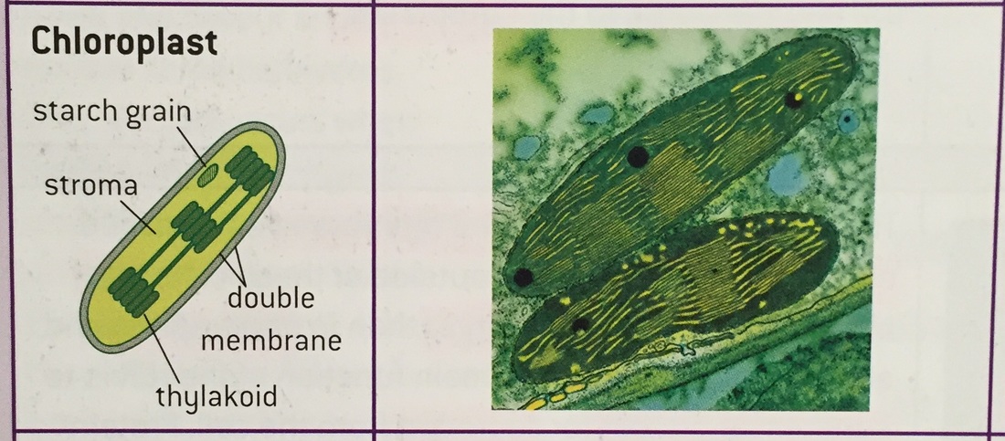
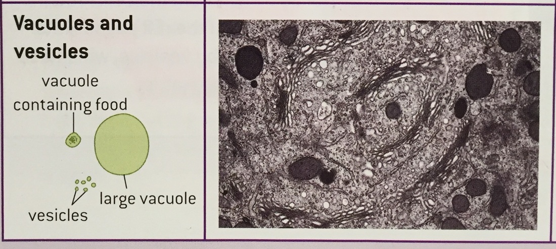
- They have all different organelles in their cell; Nucleus, Rough endoplasmic reticulum, Golgi apparatus, Lysosome, Mitochondria, Free Ribosome, Chloroplast, Vacuoles/vesicles.
- Each organelle performs a different function and has a different structure.
- There are advantages to being compartmentalised in the cell for example:
- Enzymes and substrates for a particular process can be much more concentrated than if they were spread throughout the cytoplasm.
- Substances that could cause damage to the cell can be kept inside the membrane of an organelle.
- Conditions such as pH can be maintained at an ideal level for a particular process, which may be different to the levels needed for other processes in a cell.
- Organelles with their contents can be moved around within the cell.
- Nucleus - Double membrane structure with pores through it. It contains the chromosomes consisting of DNA associated with histone proteins. Uncoiled chromosomes are spread through the nucleus and are called chromatin. This is where DNA is replicated and transcribed to form mRNA which is exported via the nuclear pores to the cytoplasm.
- Rough endoplasmic reticulum - The rER consists of flattened membrane sacs called cisternae. Attached to the outside of these cisternae are ribosomes. They are larger than in prokaryotes, 80S. They synthesise protein for secretion from the cell. Protein synthesised by the ribosomes of the rER passes into the cisternae and is then carried, by vesicles, to the Golgi apparatus by budding off.
- Golgi apparatus - The Golgi apparatus consists of flattered membrane sacs called cisternae. The cisternae are often curved and have many vesicles near by. They process proteins brought in vesicles from the rER. Most of these proteins are then carried in vesicles to the plasma membrane for secretion.
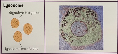
- Lysosome - Spherical, single membrane structures formed from Golgi vesicles. They contain high concentrations of protein. They contain digestive enzymes, which can be used to break down ingested food in vesicles or break down organelles in the cell or even the whole cell.
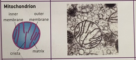
- Mitochondria - A double membrane surrounds mitochondria with the inner of these membranes invaginated to form structures called crust. The fluid inside is called the matrix. They produce ATP for the cell by aerobic cell respiration. Fat is digested here if it is being used as an energy source in the cell.
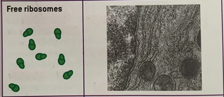
- Free ribosomes - Appears as dark granules in the cytoplasm and are not surrounded by a membrane. They are 80S. They synthesise protein, releasing it to work in the cytoplasm as enzymes or in other ways. They are constructed in a region of the nucleus called the nucleolus.
- Chloroplast - A double membrane surrounds the chloroplast. Inside are stacks of thylakoids, which are flattered sacs of membrane. They produce glucose and a wide variety of other organic compounds by photosynthesis. Starch grains may be present inside chloroplasts if they have been photosynthesising rapidly.
- Vacuoles/Vesicles - They consist of a single membrane with fluid inside. Many plant cells have large vacuoles that occupy more than half of the cell volume. Some animals absorb foods from outside and digest them inside vacuoles. Some unicellular organisms use vacuoles to expel excess water. Vesicles are very small vacuoles used to transport materials inside the cell.
How to draw a eukaryotic cell, based on an electron micrograph:
Must show - Plasma membrane, Cytoplasm with 80S ribosomes, Nucleus, Mitochondria, and all other organelles in the cell

|

|

|
|
|
|
|
|

|
|
|
|
1.3 Membrane Structure
UNDERSTANDINGS, APPLICATIONS AND SKILLS:
1.3.U1Phospholipids form bilayers in water due to the amphipathic properties of phospholipid molecules. [Amphipathic phospholipids have hydrophilic and hydrophobic properties.]
1.3.U2Membrane proteins are diverse in terms of structure, position in the membrane and function.
1.3.U3Cholesterol is a component of animal cell membranes.
1.3.A1Cholesterol in mammalian membranes reduces membrane fluidity and permeability to some solutes.
1.3.S1Drawing of the fluid mosaic model. [Drawings of the fluid mosaic model of membrane structure can be two dimensional rather than three dimensional. Individual phospholipid molecules should be shown using the symbol of a circle with two parallel lines attached. A range of membrane proteins should be shown including glycoproteins.]
1.3.S2Analysis of evidence from electron microscopy that led to the proposal of the Davson-Danielli model.
1.3.S3Analysis of the falsification of the Davson-Danielli model that led to the Singer-Nicolson model.
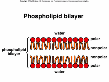
Phospholipid Bilayers
Hydrophilic - Attracted to water
Hydrophobic - NOT attracted to water
Hydrophilic - Attracted to water
Hydrophobic - NOT attracted to water
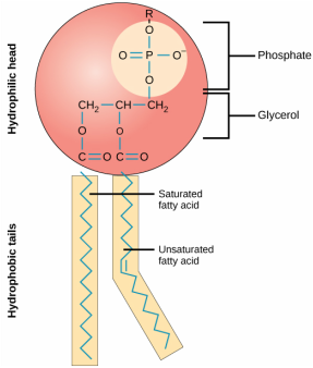
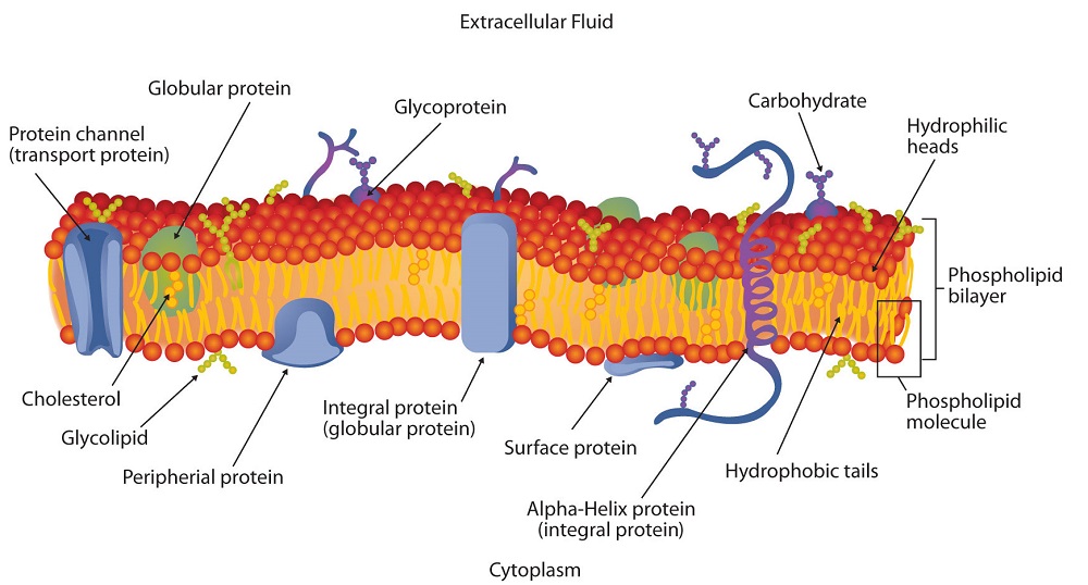
- Phospholipids are amphipathic. Meaning that parts of them are hydrophilic and parts of them are hydrophobic.
- They consist of the hydrophilic phosphate head and the hydrophobic hydrocarbon tails.
- When mixed with water they form a double layer membrane, also known as he phospholipid bilayer, because the heads are attracted to water and the tails are not.
- The heads are facing outwards and the tails are facing inwards.
- They are stable structures and form the basis of all cell membranes.
|
|
Membrane Proteins
Cell membranes have a wide range of functions including the following which make them diverse in structure and in their position.
How to draw a fluid mosaic model:
Must show - Glycoproteins, A range of other membrane proteins, Phospholipids with 2 lines attached showing the hydrocarbon chains.
Cell membranes have a wide range of functions including the following which make them diverse in structure and in their position.
- PRIMARY FUNCTION: form a barrier in which ions and hydrophilic molecules cannot pass through easily. (Carried out by the phospholipid bilayer)
- Hormones binding sites/receptors.
- Immobilised enzymes with the active site on the outside.
- Cell adhesion - to form tight junctions between groups of cells in tissues and organs.
- Cell-to-cell communication.
- Channels for passive transport to allow hydrophilic particles across by facilitated diffusion.
- Pumps for active transport.
- Integral
- Hydrophobic on at least part of their surface
- Imbedded in the hydrocarbon chains in the centre of the membrane
- Many are transmembrane, meaning they extend across the membrane with hydrophilic parts projecting through either side.
- Peripheral
- Hydrophilic on their surface.
- Mostly attached to the surface of integral proteins but the attachment is reversable.
- Some have a single hydrocarbon chain attached to them which is inserted into the membrane which anchors the protein to the membrane surface.
How to draw a fluid mosaic model:
Must show - Glycoproteins, A range of other membrane proteins, Phospholipids with 2 lines attached showing the hydrocarbon chains.

|
|
Cholesterol In Membranes
Although the two main components of membranes are phospholipids and proteins, in animal membranes they also contain cholesterol.
APPLICATION
Cholesterol in mammalian membrane reduces membrane fluidity and permeability to some solutes.
The fluidity of an animal cell membranes needs to be controlled. If it was too fluid they would be less able to control what substances pass through whereas if they were not fluid enough the movement of the cell and substances within it would be restricted. Cholesterol disrupts the regular picking go the hydrocarbon tails and prevents them crystallising and behaving as a solid. It also restricts molecule motion, therefore the fluidity of the membrane. It reduces its permeability to hydrophilic particles such as sodium ions and hydrogen ions. Its shape also helps membranes curve into a concave shape which helps lead them to the formation of vesicles during endocytosis.
Although the two main components of membranes are phospholipids and proteins, in animal membranes they also contain cholesterol.
- Its a lipid belonging to the steroids group.
- Most of the molecule is hydrophobic so it is attracted to the hydrophobic hydrocarbon tails in the membrane.
- It is positioned between phospholipids in the membrane.
APPLICATION
Cholesterol in mammalian membrane reduces membrane fluidity and permeability to some solutes.
The fluidity of an animal cell membranes needs to be controlled. If it was too fluid they would be less able to control what substances pass through whereas if they were not fluid enough the movement of the cell and substances within it would be restricted. Cholesterol disrupts the regular picking go the hydrocarbon tails and prevents them crystallising and behaving as a solid. It also restricts molecule motion, therefore the fluidity of the membrane. It reduces its permeability to hydrophilic particles such as sodium ions and hydrogen ions. Its shape also helps membranes curve into a concave shape which helps lead them to the formation of vesicles during endocytosis.
|
|
|
|
1.4 Membrane Transport
UNDERSTANDINGS, APPLICATIONS AND SKILLS:
1.4.U1Particles move across membranes by simple diffusion, facilitated diffusion, osmosis and active transport.
1.4.U2The fluidity of membranes allows materials to be taken into cells by endocytosis or released by exocytosis.
1.4.U3Vesicles move materials within cells.
1.4.A1Structure and function of sodium–potassium pumps for active transport and potassium channels for facilitated diffusion in axons.
1.4.A2Tissues or organs to be used in medical procedures must be bathed in a solution with the same osmolarity as the cytoplasm to prevent osmosis.
1.4.S1Estimation of osmolarity in tissues by bathing samples in hypotonic and hypertonic solutions. (Practical 2) [Osmosis experiments are a useful opportunity to stress the need for accurate mass and volume measurements in scientific experiments.]
1.5 The Origin Of Cells
UNDERSTANDINGS, APPLICATIONS AND SKILLS:
1.5.U1Cells can only be formed by division of pre-existing cells. [Students should be aware that the 64 codons in the genetic code have the same meanings in nearly all organisms, but that there are some minor variations that are likely to have accrued since the common origin of life on Earth.]
1.5.U2The first cells must have arisen from non-living material.
1.5.U3The origin of eukaryotic cells can be explained by the endosymbiotic theory. [Evidence for the endosymbiotic theory is expected. The origin of eukaryote cilia and flagella does not need to be included.]
1.5.A1Evidence from Pasteur’s experiments that spontaneous generation of cells and organisms does not now occur on Earth.
1.6 Cell Division
UNDERSTANDINGS, APPLICATIONS AND SKILLS:
1.6.U1Mitosis is division of the nucleus into two genetically identical daughter nuclei. [The sequence of events in the four phases of mitosis should be known. To avoid confusion in terminology, teachers are encouraged to refer to the two parts of a chromosome as sister chromatids, while they are attached to each other by a centromere in the early stages of mitosis. From anaphase onwards, when sister chromatids have separated to form individual structures, they should be referred to as chromosomes.]
1.6.U2Chromosomes condense by supercoiling during mitosis.
1.6.U3Cytokinesis occurs after mitosis and is different in plant and animal cells.
1.6.U4Interphase is a very active phase of the cell cycle with many processes occurring in the nucleus and cytoplasm.
1.6.U5Cyclins are involved in the control of the cell cycle.
1.6.U6Mutagens, oncogenes and metastasis are involved in the development of primary and secondary tumours.
1.6.A1The correlation between smoking and incidence of cancers.
1.6.S1Identification of phases of mitosis in cells viewed with a microscope or in a micrograph. [Preparation of temporary mounts of root squashes is recommended but phases in mitosis can also be viewed using permanent slides.]
1.6.S2Determination of a mitotic index from a micrograph.
Topic 2 - Molecular Biology
2.1 Molecules To Metabolism
UNDERSTANDINGS, APPLICATIONS AND SKILLS:
2.1.U1Molecular biology explains living processes in terms of the chemical substances involved.
2.1.U2Carbon atoms can form four covalent bonds allowing a diversity of stable compounds to exist.
2.1.U3Life is based on carbon compounds including carbohydrates, lipids, proteins and nucleic acids. [Sugars include monosaccharides and disaccharides. Only one saturated fat is expected and its specific name is not necessary. The variable radical of amino acids can be shown as R. The structure of individual R-groups does not need to be memorized.]
2.1.U4Metabolism is the web of all the enzyme-catalysed reactions in a cell or organism.
2.1.U5Anabolism is the synthesis of complex molecules from simpler molecules including the formation of macromolecules from monomers by condensation reactions.
2.1.U6Catabolism is the breakdown of complex molecules into simpler molecules including the hydrolysis of macromolecules into monomers.
2.1.A1Urea as an example of a compound that is produced by living organisms but can also be artificially synthesized.
2.1.S1Drawing molecular diagrams of glucose, ribose, a saturated fatty acid and a generalized amino acid. [Only the ring forms of D-ribose, alpha–D-glucose and beta-D-glucose are expected in drawings.]
2.1.S2Identification of biochemicals such as sugars, lipids or amino acids from molecular diagrams. [Students should be able to recognize from molecular diagrams that triglycerides, phospholipids and steroids are lipids. Drawings of steroids are not expected. Proteins or parts of polypeptides should be recognized from molecular diagrams showing amino acids linked by peptide bonds.]
2.2 Water
UNDERSTANDINGS, APPLICATIONS AND SKILLS:
2.2.U1Water molecules are polar and hydrogen bonds form between them.
2.2.U2Hydrogen bonding and dipolarity explain the cohesive, adhesive, thermal and solvent properties of water. [Students should know at least one example of a benefit to living organisms of each property of water. Transparency of water and maximum density at 4°C do not need to be included.]
2.2.U3Substances can be hydrophilic or hydrophobic.
2.2.A1Comparison of the thermal properties of water with those of methane. [Comparison of the thermal properties of water and methane assists in the understanding of the significance of hydrogen bonding in water.]
2.2.A2Use of water as a coolant in sweat.
2.2.A3Modes of transport of glucose, amino acids, cholesterol, fats, oxygen and sodium chloride in blood in relation to their solubility in water.
2.3 Carbohydrates And Lipids
UNDERSTANDINGS, APPLICATIONS AND SKILLS:
2.3.U1Monosaccharide monomers are linked together by condensation reactions to form disaccharides and polysaccharide polymers. [Sucrose, lactose and maltose should be included as examples of disaccharides produced by combining monosaccharides. The structure of starch should include amylose and amylopectin.]
2.3.U2Fatty acids can be saturated, monounsaturated or polyunsaturated. [Named examples of fatty acids are not required.]
2.3.U3Unsaturated fatty acids can be cis or trans isomers.
2.3.U4Triglycerides are formed by condensation from three fatty acids and one glycerol.
2.3.A1Structure and function of cellulose and starch in plants and glycogen in humans.
2.3.A2Scientific evidence for health risks of trans fats and saturated fatty acids.
2.3.A3Lipids are more suitable for long-term energy storage in humans than carbohydrates.
2.3.A4Evaluation of evidence and the methods used to obtain the evidence for health claims made about lipids.
2.3.S1Use of molecular visualization software to compare cellulose, starch and glycogen.
2.3.S2Determination of body mass index by calculation or use of a nomogram.
2.4 Proteins
UNDERSTANDINGS, APPLICATIONS AND SKILLS:
2.4.U1Amino acids are linked together by condensation to form polypeptides.
2.4.U2There are 20 different amino acids in polypeptides synthesized on ribosomes. [Students should know that most organisms use the same 20 amino acids in the same genetic code although there are some exceptions. Specific examples could be used for illustration.]
2.4.U3Amino acids can be linked together in any sequence giving a huge range of possible polypeptides.
2.4.U4The amino acid sequence of polypeptides is coded for by genes.
2.4.U5A protein may consist of a single polypeptide or more than one polypeptide linked together.
2.4.U6The amino acid sequence determines the three-dimensional conformation of a protein.
2.4.U7Living organisms synthesize many different proteins with a wide range of functions.
2.4.U8Every individual has a unique proteome.
2.4.A1Rubisco, insulin, immunoglobulins, rhodopsin, collagen and spider silk as examples of the range of protein functions. [The detailed structure of the six proteins selected to illustrate the functions of proteins is not needed.]
2.4.A2Denaturation of proteins by heat or by deviation of pH from the optimum. [Egg white or albumin solutions can be used in denaturation experiments.]
2.4.S1Drawing molecular diagrams to show the formation of a peptide bond.
2.5 Enzymes
UNDERSTANDINGS, APPLICATIONS AND SKILLS:
2.5.U1Enzymes have an active site to which specific substrates bind.
2.5.U2Enzyme catalysis involves molecular motion and the collision of substrates with the active site.
2.5.U3Temperature, pH and substrate concentration affect the rate of activity of enzymes. [Students should be able to sketch graphs to show the expected effects of temperature, pH and substrate concentration on the activity of enzymes. They should be able to explain the patterns or trends apparent in these graphs.]
2.5.U4Enzymes can be denatured.
2.5.U5Immobilized enzymes are widely used in industry.
2.5.A1Methods of production of lactose-free milk and its advantages. [Lactase can be immobilized in alginate beads and experiments can then be carried out in which the lactose in milk is hydrolysed.]
2.5.S1Design of experiments to test the effect of temperature, pH and substrate concentration on the activity of enzymes.
2.5.S2Experimental investigation of a factor affecting enzyme activity. (Practical 3)
2.6 Structure Of DNA And RNA
UNDERSTANDINGS, APPLICATIONS AND SKILLS:
2.6.U1The nucleic acids DNA and RNA are polymers of nucleotides.
2.6.U2DNA differs from RNA in the number of strands present, the base composition and the type of pentose.
2.6.U3DNA is a double helix made of two antiparallel strands of nucleotides linked by hydrogen bonding between complementary base pairs.
2.6.A1Crick and Watson’s elucidation of the structure of DNA using model making.
2.6.S1Drawing simple diagrams of the structure of single nucleotides of DNA and RNA, using circles, pentagons and rectangles to represent phosphates, pentoses and bases. [In diagrams of DNA structure, the helical shape does not need to be shown, but the two strands should be shown antiparallel. Adenine should be shown paired with thymine and guanine with cytosine, but the relative lengths of the purine and pyrimidine bases do not need to be recalled, nor the numbers of hydrogen bonds between the base pairs.]
2.7 DNA Replication, Transcription And Translation
UNDERSTANDINGS, APPLICATIONS AND SKILLS:
2.7.U1The replication of DNA is semi-conservative and depends on complementary base pairing.
2.7.U2Helicase unwinds the double helix and separates the two strands by breaking hydrogen bonds.
2.7.U3DNA polymerase links nucleotides together to form a new strand, using the pre-existing strand as a template. [The different types of DNA polymerase do not need to be distinguished.]
2.7.U4Transcription is the synthesis of mRNA copied from the DNA base sequences by RNA polymerase.
2.7.U5Translation is the synthesis of polypeptides on ribosomes.
2.7.U6The amino acid sequence of polypeptides is determined by mRNA according to the genetic code.
2.7.U7Codons of three bases on mRNA correspond to one amino acid in a polypeptide.
2.7.U8Translation depends on complementary base pairing between codons on mRNA and anticodons on tRNA.
2.7.A1Use of Taq DNA polymerase to produce multiple copies of DNA rapidly by the polymerase chain reaction (PCR).
2.7.A2Production of human insulin in bacteria as an example of the universality of the genetic code allowing gene transfer between species.
2.7.S1Use a table of the genetic code to deduce which codon(s) corresponds to which amino acid.
2.7.S2Analysis of Meselson and Stahl’s results to obtain support for the theory of semi-conservative replication of DNA.
2.7.S3Use a table of mRNA codons and their corresponding amino acids to deduce the sequence of amino acids coded by a short mRNA strand of known base sequence.
2.7.S4Deducing the DNA base sequence for the mRNA strand.
2.8 Cell Respiration
UNDERSTANDINGS, APPLICATIONS AND SKILLS:
2.8.U1Cell respiration is the controlled release of energy from organic compounds to produce ATP. [Details of the metabolic pathways of cell respiration are not needed but the substrates and final waste products should be known.]
2.8.U2ATP from cell respiration is immediately available as a source of energy in the cell.
2.8.U3Anaerobic cell respiration gives a small yield of ATP from glucose.
2.8.U4Aerobic cell respiration requires oxygen and gives a large yield of ATP from glucose.
2.8.A1Use of anaerobic cell respiration in yeasts to produce ethanol and carbon dioxide in baking.
2.8.A2Lactate production in humans when anaerobic respiration is used to maximize the power of muscle contractions.
2.8.S1Analysis of results from experiments involving measurement of respiration rates in germinating seeds or invertebrates using a respirometer. [There are many simple respirometers which could be used. Students are expected to know that an alkali is used to absorb CO2, so reductions in volume are due to oxygen use. Temperature should be kept constant to avoid volume changes due to temperature fluctuations.]
2.9 Photosynthesis
UNDERSTANDINGS, APPLICATIONS AND SKILLS:
2.9.U1Photosynthesis is the production of carbon compounds in cells using light energy.
2.9.U2Visible light has a range of wavelengths with violet the shortest wavelength and red the longest.
2.9.U3Chlorophyll absorbs red and blue light most effectively and reflects green light more than other colours. [Students should know that visible light has wavelengths between 400 and 700 nanometres, but they are not expected to recall the wavelengths of specific colours of light.]
2.9.U4Oxygen is produced in photosynthesis from the photolysis of water.
2.9.U5Energy is needed to produce carbohydrates and other carbon compounds from carbon dioxide.
2.9.U6Temperature, light intensity and carbon dioxide concentration are possible limiting factors on the rate of photosynthesis.
2.9.A1Changes to the Earth’s atmosphere, oceans and rock deposition due to photosynthesis.
2.9.S1Drawing an absorption spectrum for chlorophyll and an action spectrum for photosynthesis.
2.9.S2Design of experiments to investigate the effect of limiting factors on photosynthesis. [Water free of dissolved carbon dioxide for photosynthesis experiments can be produced by boiling and cooling water.]
2.9.S3Separation of photosynthetic pigments by chromatograph. (Practical 4) [Paper chromatography can be used to separate photosynthetic pigments but thin layer chromatography gives better results.]
Topic 3 - Genetics
3.1 Genes
UNDERSTANDINGS, APPLICATIONS AND SKILLS:
3.1.U1A gene is a heritable factor that consists of a length of DNA and influences a specific characteristic.
3.1.U2A gene occupies a specific position on a chromosome.
3.1.U3The various specific forms of a gene are alleles.
3.1.U4Alleles differ from each other by one or only a few bases.
3.1.U5New alleles are formed by mutation. [Deletions, insertions and frame shift mutations do not need to be included.]
3.1.U6The genome is the whole of the genetic information of an organism.
3.1.U7The entire base sequence of human genes was sequenced in the Human Genome Project.
3.1.A1The causes of sickle cell anemia, including a base substitution mutation, a change to the base sequence of mRNA transcribed from it and a change to the sequence of a polypeptide in hemoglobin. [Students should be able to recall one specific base substitution that causes glutamic acid to be substituted by valine as the sixth amino acid in the hemoglobin polypeptide.]
3.1.A2Comparison of the number of genes in humans with other species. [The number of genes in a species should not be referred to as genome size as this term is used for the total amount of DNA. At least one plant and one bacterium should be included in the comparison and at least one species with more genes and one with fewer genes than a human.]
3.1.S1Use of a database to determine differences in the base sequence of a gene in two species. [The Genbank® database can be used to search for DNA base sequences. The cytochrome C gene sequence is available for many different organisms and is of particular interest because of its use in reclassifying organisms into three domains.]
Genes
- A gene is a heritable factor that consists of a much shorted length if DNA than a chromosomes.
- Chromosomes carry may genes.
- Each gene occupies a specific position on the type of chromosomes (e.g. chromosome 5) where it's located.
- This position is called the locus of the gene.
- Alleles are pairs of heritable factors that are alternative forms of the same gene.
- There can be more than two alleles of a gene.
- As they are alternative forms of the same gene, they occupy the same position on one type of chromosome, same locus. But only one allele can occupy the locus of the gene on a chromosome. SO we can expect two copies of a gene to be present. This could be two of the same allele or two different alleles.
- A gene consists of a length of DNA, with a base sequence that can be hundreds or thousands of bases long.
- The different alleles of a gene have slight variations in the base sequence. Usually one or a very small number of bases are different.
- Positions in a gene where more than one base may be present are called single nucleotide polymorphisms (SNPs), pronounced snips. Several snips can be present in a gene. But even in this case, the alleles of the gene differ by only a few bases.
- New alleles are formed from other alleles by gene mutation.
- Mutations are random changes. The most significant type of mutation is a base substitution.
- A random change to an allele that has developed by evolution over millions of years is unlikely to be beneficial. Almost all mutations are therefore either neutral or harmful.
- Some mutations are lethal - cause the death of the cell.
- Mutations in body cells are eliminated when the individual dies, but mutations in cells that develop into gametes can be passes on to offspring and cause genetic disease.
Genome
- The genome is that whole of the genetic information of an organism.
- Genetic information is contained in DNA, so a living organism's genome is the entire base sequence of each of its DNA molecules.
- Humans genome consists of 46 molecules that form the chromosomes in the nucleus + the DNA molecules in the mitochondria.
- This is the same in other animals apart from the number of chromosomes may vary
- Plants genome consists of the DNA molecules of chromosomes in the nucleus + the DNA molecules in the mitochondria and chloroplast
- The genome of prokaryotes is much smaller and consists of DNA in the circular chromosome + any plasmids that are present.
- Humans genome consists of 46 molecules that form the chromosomes in the nucleus + the DNA molecules in the mitochondria.
All about the human genome
|
|
|
|
3.2 Chromosomes
UNDERSTANDINGS, APPLICATIONS AND SKILLS:
3.2.U1Prokaryotes have one chromosome consisting of a circular DNA molecule.
3.2.U2Some prokaryotes also have plasmids but eukaryotes do not.
3.2.U3Eukaryote chromosomes are linear DNA molecules associated with histone proteins.
3.2.U4In a eukaryote species there are different chromosomes that carry different genes.
3.2.U5Homologous chromosomes carry the same sequence of genes but not necessarily the same alleles of those genes.
3.2.U6Diploid nuclei have pairs of homologous chromosomes.
3.2.U7Haploid nuclei have one chromosome of each pair. [The two DNA molecules formed by DNA replication prior to cell division are considered to be sister chromatids until the splitting of the centromere at the start of anaphase. After this, they are individual chromosomes.]
3.2.U8The number of chromosomes is a characteristic feature of members of a species.
3.2.U9A karyogram shows the chromosomes of an organism in homologous pairs of decreasing length. [The terms karyotype and karyogram have different meanings. Karyotype is a property of a cell—the number and type of chromosomes present in the nucleus, not a photograph or diagram of them.]
3.2.U10Sex is determined by sex chromosomes and autosomes are chromosomes that do not determine sex.
3.2.A1Cairns’ technique for measuring the length of DNA molecules by autoradiography.
3.2.A2Comparison of genome size in T2 phage,Escherichia coli, Drosophila melanogaster, Homo sapiens and Paris japonica. [Genome size is the total length of DNA in an organism. The examples of genome and chromosome number have been selected to allow points of interest to be raised.]
3.2.A3Comparison of diploid chromosome numbers of Homo sapiens, Pan troglodytes, Canis familiaris, Oryza sativa, Parascaris equorum.
3.2.A4Use of karyograms to deduce sex and diagnose Down syndrome in humans.
3.2.S1Use of databases to identify the locus of a human gene and its polypeptide product.
3.3 Meiosis
UNDERSTANDINGS, APPLICATIONS AND SKILLS:
3.3.U1One diploid nucleus divides by meiosis to produce four haploid nuclei.
3.3.U2The halving of the chromosome number allows a sexual life cycle with fusion of gametes.
3.3.U3DNA is replicated before meiosis so that all chromosomes consist of two sister chromatids.
3.3.U4The early stages of meiosis involve pairing of homologous chromosomes and crossing over followed by condensation. [The process of chiasmata formation need not be explained.]
3.3.U5Orientation of pairs of homologous chromosomes prior to separation is random.
3.3.U6Separation of pairs of homologous chromosomes in the first division of meiosis halves the chromosome number.
3.3.U7Crossing over and random orientation promotes genetic variation.
3.3.U8Fusion of gametes from different parents promotes genetic variation.
3.3.A1Non-disjunction can cause Down syndrome and other chromosome abnormalities.
3.3.A2Studies showing age of parents influences chances of non-disjunction.
3.3.A3Description of methods used to obtain cells for karyotype analysis e.g. chorionic villus sampling and amniocentesis and the associated risks.
3.3.S1Drawing diagrams to show the stages of meiosis resulting in the formation of four haploid cells. [Drawings of the stages of meiosis do not need to include chiasmata. Preparation of microscope slides showing meiosis is challenging and permanent slides should be available in case no cells in meiosis are visible in temporary mounts.]
3.4 Inheritance
UNDERSTANDINGS, APPLICATIONS AND SKILLS:
3.4.U1Mendel discovered the principles of inheritance with experiments in which large numbers of pea plants were crossed.
3.4.U2Gametes are haploid so contain only one allele of each gene.
3.4.U3The two alleles of each gene separate into different haploid daughter nuclei during meiosis.
3.4.U4Fusion of gametes results in diploid zygotes with two alleles of each gene that may be the same allele or different alleles.
3.4.U5Dominant alleles mask the effects of recessive alleles but co-dominant alleles have joint effects.
3.4.U6Many genetic diseases in humans are due to recessive alleles of autosomal genes, although some genetic diseases are due to dominant or co-dominant alleles.
3.4.U7Some genetic diseases are sex-linked. The pattern of inheritance is different with sex-linked genes due to their location on sex chromosomes. [Alleles carried on X chromosomes should be shown as superscript letters on an upper case X, such as Xh.]
3.4.U8Many genetic diseases have been identified in humans but most are very rare.
3.4.U9Radiation and mutagenic chemicals increase the mutation rate and can cause genetic diseases and cancer.
3.4.A1Inheritance of ABO blood groups. [The expected notation for ABO blood group alleles: O = i, A=IA, B = IB.]
3.4.A2Red-green colour blindness and hemophilia as examples of sex-linked inheritance.
3.4.A3Inheritance of cystic fibrosis and Huntington’s disease.
3.4.A4Consequences of radiation after nuclear bombing of Hiroshima and accident at Chernobyl.
3.4.S1Construction of Punnett grids for predicting the outcomes of monohybrid genetic crosses.
3.4.S2Comparison of predicted and actual outcomes of genetic crosses using real data.
3.4.S3Analysis of pedigree charts to deduce the pattern of inheritance of genetic diseases.
3.5 Genetic Modification And Biotechnology
Topic 7 - Nucleic Acids
UNDERSTANDINGS, APPLICATIONS AND SKILLS:
3.5.U1Gel electrophoresis is used to separate proteins or fragments of DNA according to size.
3.5.U2PCR can be used to amplify small amounts of DNA.
3.5.U3DNA profiling involves comparison of DNA.
3.5.U4Genetic modification is carried out by gene transfer between species.
3.5.U5Clones are groups of genetically identical organisms, derived from a single original parent cell.
3.5.U6Many plant species and some animal species have natural methods of cloning.
3.5.U7Animals can be cloned at the embryo stage by breaking up the embryo into more than one group of cells.
3.5.U8Methods have been developed for cloning adult animals using differentiated cells.
3.5.A1Use of DNA profiling in paternity and forensic investigations.
3.5.A2Gene transfer to bacteria using plasmids makes use of restriction endonucleases and DNA ligase.
3.5.A3Assessment of the potential risks and benefits associated with genetic modification of crops.
3.5.A4Production of cloned embryos produced by somatic-cell nuclear transfer. [Dolly can be used as an example of somatic-cell transfer.]
3.5.S1Design of an experiment to assess one factor affecting the rooting of stem-cuttings. [A plant species should be chosen for rooting experiments that forms roots readily in water or a solid medium.]
3.5.S2Analysis of examples of DNA profiles. [Students should be able to deduce whether or not a man could be the father of a child from the pattern of bands on a DNA profile.]
3.5.S3Analysis of data on risks to monarch butterflies of Bt crops.
7.1 DNA Structure And Replication
UNDERSTANDINGS, APPLICATIONS AND SKILLS:
7.1.U1Nucleosomes help to supercoil the DNA.
7.1.U2DNA structure suggested a mechanism for DNA replication.
7.1.U3DNA polymerases can only add nucleotides to the 3’ end of a primer.
7.1.U4DNA replication is continuous on the leading strand and discontinuous on the lagging strand. [Details of DNA replication differ between prokaryotes and eukaryotes. Only the prokaryotic system is expected.]
7.1.U5DNA replication is carried out by a complex system of enzymes. [The proteins and enzymes involved in DNA replication should include helicase, DNA gyrase, single strand binding proteins, DNA primase and DNA polymerases I and III.]
7.1.U6Some regions of DNA do not code for proteins but have other important functions. [The regions of DNA that do not code for proteins should be limited to regulators of gene expression, introns, telomeres and genes for tRNAs.]
7.1.A1Rosalind Franklin’s and Maurice Wilkins’ investigation of DNA structure by X-ray diffraction.
7.1.A2Use of nucleotides containing dideoxyribonucleic acid to stop DNA replication in preparation of samples for base sequencing.
7.1.A3Tandem repeats are used in DNA profiling.
7.1.S1Analysis of results of the Hershey and Chase experiment providing evidence that DNA is the genetic material.
7.1.S2Utilization of molecular visualization software to analyse the association between protein and DNA within a nucleosome.
7.2 Transcription And Gene Expression
UNDERSTANDINGS, APPLICATIONS AND SKILLS:
7.2.U1Transcription occurs in a 5’ to 3’ direction. [RNA polymerase adds the 5´ end of the free RNA nucleotide to the 3´ end of the growing mRNA molecule.]
7.2.U2Nucleosomes help to regulate transcription in eukaryotes.
7.2.U3Eukaryotic cells modify mRNA after transcription.
7.2.U4Splicing of mRNA increases the number of different proteins an organism can produce.
7.2.U5Gene expression is regulated by proteins that bind to specific base sequences in DNA.
7.2.U6The environment of a cell and of an organism has an impact on gene expression.
7.2.A1The promoter as an example of non-coding DNA with a function.
7.2.S1Analysis of changes in the DNA methylation patterns.
7.3 Translation
UNDERSTANDINGS, APPLICATIONS AND SKILLS:
7.3.U1Initiation of translation involves assembly of the components that carry out the process. [Examples of start codons are not required. Names of the tRNA binding sites are expected as well as their roles.]
7.3.U2Synthesis of the polypeptide involves a repeated cycle of events.
7.3.U3Disassembly of the components follows termination of translation. [Examples of stop codons are not required.]
7.3.U4Free ribosomes synthesize proteins for use primarily within the cell.
7.3.U5Bound ribosomes synthesize proteins primarily for secretion or for use in lysosomes.
7.3.U6Translation can occur immediately after transcription in prokaryotes due to the absence of a nuclear membrane.
7.3.U7The sequence and number of amino acids in the polypeptide is the primary structure.
7.3.U8The secondary structure is the formation of alpha helices and beta pleated sheets stabilized by hydrogen bonding.
7.3.U9The tertiary structure is the further folding of the polypeptide stabilized by interactions between R groups. [Polar and non-polar amino acids are relevant to the bonds formed between R groups.]
7.3.U10The quaternary structure exists in proteins with more than one polypeptide chain. [Quaternary structure may involve the binding of a prosthetic group to form a conjugated protein.]
7.3.A1tRNA-activating enzymes illustrate enzyme–substrate specificity and the role of phosphorylation.
7.3.S1Identification of polysomes in electron micrographs of prokaryotes and eukaryotes.
7.3.S2The use of molecular visualization software to analyse the structure of eukaryotic ribosomes and a tRNA molecule.
